Menu

The study titled, ‘Root Resorption: Causes and Remedies’ was originally published by Dentistry Today.
To successfully treat root resorption can be a challenge for many clinicians. This is due to the complex nature of the diagnosis. This article aims to cover the main causes of root resorption followed by a treatment plan. Let us first begin by having a brief understanding of root resorption and its types.
What is Root Resorption?
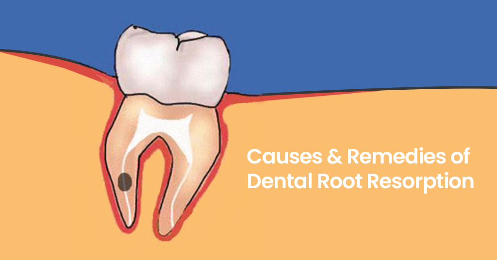
The progressive loss of cementum and dentin due to the action of odontoclasts is known as root resorption (RR). It can occur as a pathological or a physiological phenomenon and it presents in various forms.
There can be external root resorption (ERR) that usually appears on the side of the root. For instance:
There can also be internal root resorption (IRR) that appears first within the root canal.
RR can affect the entire root or transpire in any third of the root. It is crucial to detect if RR is IRR or ERR. The reason being, a wrong diagnosis can lead to an ineffective treatment plan. The most identifiable cause of RR is usually trauma. You can clearly tell if the RR is external or internal by preparing a limited CBCT image.
Causes of Adult Root Resorption
EPR can affect the root surface and teeth that have chronic apical periodontitis. It can quickly advance to a point where a complete root surface can be resorbed in a couple of months if it is not treated. Orthodontic tooth movement is one of the most usual causes of RR. If orthodontic pressures are too strong for the patient to endure, it could trigger an internal and external generalized RR. Oral trauma due to automobile accidents, fighting, and athletic injuries can cause RR as well.
The clinical presentation of ankylosis or RER is quite distinct. For instance, there will be a high pitched sound when the tooth is percussed with a mouth mirror handle. After a radiographic image, the clinician will notice three-fourths of the root that can be replaced by a bone. The clinician will also notice that the crown will be quite stable when checked for mobility. Even after the root has been replaced with bone, the tooth will stabilize and function well for years to come.
Some other causes for RR could be chemotherapy, heating a tooth beyond a certain point during internal bleaching of a crown, and avulsed teeth replantation.
Repairing Root Resorption
Quite a few studies have shown that calcium silicates can be considered for arresting RR. For instance, where access can be gained in the resorptive defect, the medical professional must mix Biodentine and must use an amalgam carrier to carry it to the resorptive defect. Since the initial setting happens between 10-12 mins after mixing, the Biodentine can be carved within a couple of minutes. The biocompatible quality and easy handling of the Biodentine makes it an excellent restorative material for RR.
Conclusion
The clinician must have a heightened suspicion about the occurrence of a RR when the patient has had oral trauma due to an automobile accident, athletic injuries and even fights. RR must also be considered in cases of aggressive orthodontic treatment. After careful clinical testing, the patient must return after 2 weeks to repeat the tests. If the results of both the tests are similar, then request the patient to return in 6 weeks and compare the patient’s current findings to the first test. If there is no change, then the patient must be advised for a follow-up test every 3 months for 2 years.


| PRODUCTS | QTY | PRICE | VALUE in INR |
|---|
| PRODUCTS | QTY | PRICE | VALUE in INR |
|---|