Menu

The study titled ‘Reassessing Current Endodontic Treatment’ was originally published by Dentistry Today.
The current advancements in radiography, CBCT and, apex locators have enabled dentists to have a better understanding of treating root canal morphologies. Yet, even with the advancements in endodontic canal preparation, the following are not consistently addressed:
Let us look at the challenges that dentists face with current endodontic treatment.
Challenges with Current Endodontic Treatment
While providing endodontic treatment, clinicians cannot directly observe the complicated root anatomy they are treating. Instead, they need to depend on a 2-D radiograph that represents a 3-D biological canal. Even with the use of CBCT in endodontics that displays a 3-D canal image, the canal morphology that is revealed by the CBCT still proves to be a clinical challenge. It is difficult to chemomechanical prepare a root canal system.
What’s more, dentists are overwhelmed with the latest rotary NiTi file systems. Endodontic file manufacturers are finding ways to appeal to the dentist’s clinical needs and ensure canal preparation is effective, simple, and uses the least number of files. However, the new files have a flaw. They lack the ability to anatomically prepare canals. Hence, files still cannot solve the basic problem of debriding a root canal system.
The reason being, when rotating, the NiTi rotary files stay centered in the canal. So, they can’t contact the dentinal walls that are filled with irregularities or invaginations. In a recent study, it was found that endodontic failures occurred due to the persistence of bacterial infection in the canal space.
Along with the physical morphological challenges clinicians face in preparing a canal, there are many quantitative challenges as well. It is quite common for dentists to struggle during endodontic treatment to maintain and determine proper working length through the canal preparation. It is also difficult to determine the right apical size, in order to enlarge a canal.
The question then arises:
How can Dentists Prepare for a Root Canal?
To find the solution, dentists need to first think like a physician. For instance, if a physician was to provide root canal treatment, they would ignore quantitative assessments that dentists usually become obsessed about.
Physicians would look at the situation more qualitatively. While providing endodontic treatment, a physician would extract the infected or inflamed pulpal tissue while preserving the natural canal morphology. They would not be assessing the canal diameter tapers as dentists would. Finally, they would also think about providing endodontic treatment from a biological approach.
BIOACTIVE ENDODONTICS
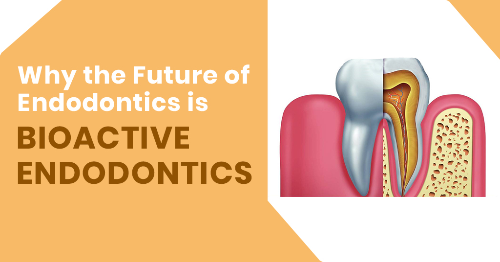
It is vital for dentists to know that the future of endodontics is bioactive endodontics. Bioactive is defined as having a biological effect. To achieve bioactive endodontic treatment, either regenerative endodontics or vital pulp cryotherapy must be carried out.
1) Vital Pulp Cryotherapy
In vital pulp cryotherapy, sterile ice is used in conjunction with bioceramics, EDTA, and restorative materials on the pulp tissue that’s exposed directly or indirectly because of a carious lesion.
Vital pulp cryotherapy is usually carried on teeth that require a partial pulpectomy or a pulp cap that can later be restored with composite restoration or an amalgam. This technique is contraindicated when the tooth has any one of the following:
If it is not possible for the tooth to be permanently restored with an amalgam or composite, this technique can be contraindicated, and the tooth must be treated with a complete pulpectomy.
2) Regenerative Endodontics
Regenerative endodontics has proven to be a biological approach to endodontic treatment compared to the current clinical methodology. This technique can be done on necrotic or vital pulps of mature and immature teeth. In order to fill a prepared canal, regenerative endodontics makes use of periradicular blood. This eliminates the need for cold lateral compaction techniques and warm vertical. It also eliminates the use of carrier-based root-filling materials that are needed for canal obturation.
After a regenerative endodontic procedure, the generated tissues in the canals are bone-like, periodontal ligament-like, and cementum-like tissues with nerves and blood vessels. However, these tissues cannot be classified as true pulpal tissue. As opposed to foreign obturation materials, they are the host’s vital tissue.
Listed below are some canal preparation guidelines that need to be considered before performing regenerative endodontics.
Conclusion
To summarize, bioactive endodontics is the future of endodontics. The treatment can be achieved by regenerative endodontics or vital pulp cryotherapy. In vital pulp cryotherapy, sterile ice is used to reduce pulpal inflammation. In regenerative endodontics, the dentist needs to first undertake canal preparation. This is followed by bleeding induction, from the periapical area into the canal.
Source:
https://www.dentistrytoday.com/articles/10711


| PRODUCTS | QTY | PRICE | VALUE in INR |
|---|
| PRODUCTS | QTY | PRICE | VALUE in INR |
|---|